Face pareidolia

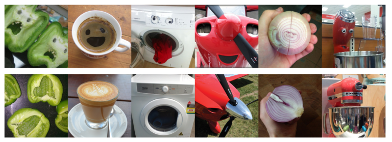
Face pareidolia is the perception of illusory faces in inanimate objects. It’s a specific example of the more general phenomenon known as pareidolia, which is the perception of any meaningful structure that isn’t actually there (e.g. seeing a cloud in the shape of another object such as a lion).
Why study face pareidolia? Our visual system is generally optimized for quickly detecting faces, but a side effect of this hypersensitivity to faces is that we sometimes see faces where there are none. In this way face pareidolia can be thought of as a mistake of face detection. Visual illusions or errors of perception are often informative about how our brain works, because they can reveal its biases and assumptions. So in addition to face pareidolia being an interesting phenomenon to study in itself, it is also a useful tool for understanding how the human brain processes faces more generally.
How does our brain process face pareidolia? Examples of face pareidolia are interesting from a cognitive neuroscience perspective because they are both a face and an object. There are regions in the human brain that preferentially respond to faces such as the fusiform face area (FFA) and the occipital face area (OFA), and other areas such as the lateral occipital cortex (LO) that are sensitive to the properties of objects such as their shape. Since examples of face pareidolia are a special case of a face in an object, we wanted to find out how illusory faces in objects were represented in the human brain.

In a multimodal neuroimaging study (Wardle et al., 2020, Nature Communications), we measured the brain activity of participants using functional magnetic resonance imaging (fMRI) and magnetoencephalography (MEG) while they looked at images of human faces, face pareidolia, and ordinary objects that were matched as closely as possible to the examples of face pareidolia, but did not have a face. fMRI has much higher spatial resolution, and MEG has much higher temporal resolution, so we had the advantages of both neuroimaging methods.

Example stimuli from Wardle et al. (2020): face pareidolia and matched objects without a face.

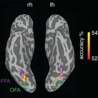
Searchlight decoding of illusory faces versus matched objects from fMRI data.
First, we used the fMRI data to identify which regions of the brain respond to illusory faces. We used a decoding approach (machine learning) to train a classifier to distinguish between patterns of brain activity measured with fMRI associated with viewing examples of face pareidolia versus viewing similar objects without a face. We then tested the ability of the classifier to decide whether someone was looking at an illusory face or a similar object without a face based only on their patterns of brain activity.
Only patterns of brain activity in the face-selective regions (fusiform face area and occipital face area) were informative about whether someone was currently looking at an illusory face in an object or not.
The evolution of the brain’s representation of illusory faces over time as measured with MEG.
Next, we used the whole-brain MEG data to identify how the representation of illusory faces evolves over time.
The movie to the left is a schematic representation (produced by multidimensional scaling) of the brain’s representation of human faces, illusory faces, and matched objects over time. Each colored dot represents one particular image out of the 96 images we used in the experiment. Dots that are close together represent images that have similar brain representations; dots that are far apart represent images that have dissimilar brain representations.
key features:
0 ms: the image is shown to the participant (for 200 ms)
130 ms: some illusory faces are similar to their matched objects, some are similar to human faces
160 ms: illusory faces are most distinct from both human faces and objects
260 ms: illusory faces are now represented most similarly to objects, not faces.
Although illusory faces were initially represented as more similar to human faces than objects were, this representation rapidly evolved in just a quarter of a second so that illusory faces were represented more like objects than faces.
Conclusions
- The erroneous face-like response to illusory faces is confined to face-selective cortical areas and is incredibly brief.
- Although illusory faces are initially more similar to faces in the brain’s representation than other objects are, within a quarter of a second this representation transforms and they are represented like objects.
- Illusory faces are represented uniquely in the human brain compared to real faces and ordinary objects.
- Our visual system is optimized for the rapid detection of face-like structure, broadly defined. This is adaptive as a false positive is usually less costly than a missed face.
Citation: Wardle, S. G., Taubert, J., Teichmann, L., & Baker, C. I. (2020). Rapid and dynamic processing of face pareidolia in the human brain. Nature Communications, 11, 4518. [link]